Information Technology in Digital Pathology
The future paradigm of Pathology will be Digital. Instead of conventional microscopy, a pathologist will perform diagnosis through interaction with images on computer screens and performing quantitative analysis. Advances in digital pathology are generating huge volumes of whole slide images which are providing opportunities for developing and evaluating new and more effective treatments that may revolutionize care of patients. The challenge is to apply information technology and image analysis methods to exploit the new and emerging digital pathology technologies effectively in order to process and model all the data.
Targeting industry and academic experts working in Digital Pathology, this workshop will provide the opportunity to generate and discuss different analysis methods, case study examples and procedures in this field. The ‘IT in Digital Pathology’ workshop will discuss the following topics:
In recent years, digital imaging has been applied to many medical fields. Thanks to technological improvements of hardware and software, digital microscopy has become an important diagnostic tool in surgical pathology. Digital microscopy creates the digital version of the whole glass slides (Whole Slide Image – WSI), which can be dynamically viewed, navigated and magnified on a computer monitor across computer networks. Digital slides can be integrated into existing hospital databases and accessed through intranet or the Internet for teaching, primary diagnosis, teleconsultation and quality assurance. New technologies and the state of the art in digital pathology will be discussed in Section 1: ‘Strategy and Technology Digital Pathology’.
To make medical decisions, healthcare professionals require that all necessary information is both correct and easily available. Collaborative Digital Anatomic Pathology refers to the use of information technology that supports the creation and sharing or exchange of information, including metadata and images, during the complex workflow followed in an Anatomic Pathology department from specimen reception to report transmission and exploitation. Collaborative Digital Anatomic Pathology is supported by standardization efforts towards interoperability and knowledge representation for shareable and computable clinical information. These efforts will be discussed in Section 2: ‘Standards and Specifications in Pathology’.
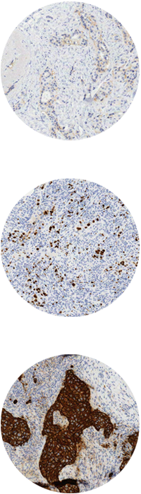
WSI is already impacting pathology practices. Digital images allow applying the digital algorithms for a variety of analysis and quantification processes. The advances in diagnosis using WSI processing tools and emerging research in this area will be the topics for Section 3: ‘Image Analysis in Digital Pathology’ and Section 4: ‘Emerging Research’, respectively.
The workshop is free, there is a cost of 35€ to cover meals. This may be paid by PayPal at grupo.visilab@uclm.es or on your arrival. Please note that registration is compulsory.
Certificates of attendance will be issued as of Tuesday, 26th May only to those previously registered.
register